The microscopy of vegetable foods, with special reference to the detection of adulteration and the diagnosis of mixtures (1916)
Winton, Andrew Lincoln, 1864-1946, Moeller, Josef, 1848-1924, Winton, Kate Grace Barber, 1882- · New York, John Wiley & sons, inc.; [etc., etc.] · leaf 536
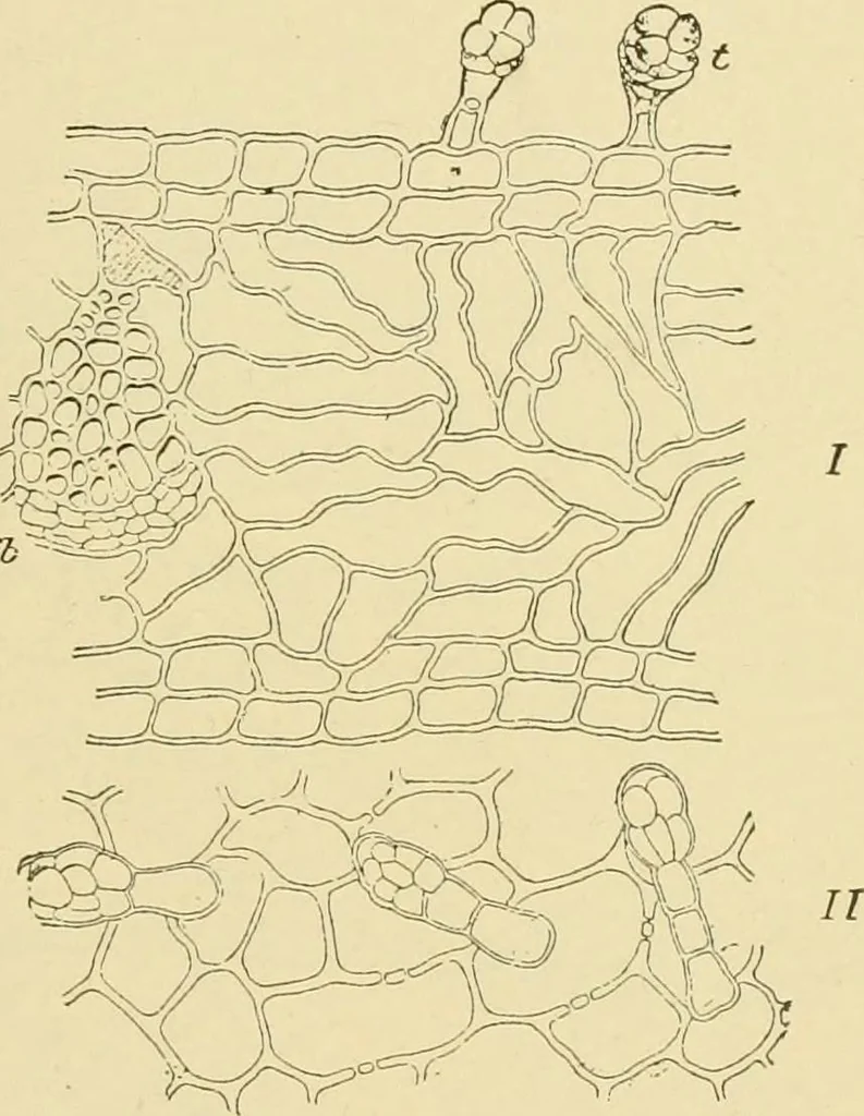
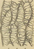
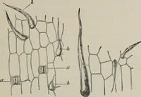
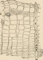
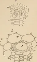
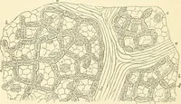
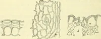
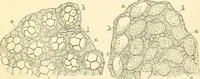
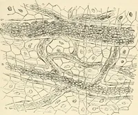
KA. 517 Calyx. Sections of the dry material swell considerably in water, dis-playing an uncommonly large-celled tissue. 1. The Outer Epidermis (Fig. 433), as seen in surface view, consistsof large, flat, moderately thick-walled, sharply-polygonal cells, with a fewstomata, 2. Mesophyl (Fig. 434).… … Fig. 434. Paprika. / cross section of calyx showing / hairs of inner (upper) epidermisand g}b fibro-vascular bundle. // inner (upper) epidermis of calyx, in surface view.(TsCHiRCH and Oesterle.) waLled, forming a spongy parenchyma. Through the inner layers runthe fibro-vascular bundles. Chlorophyl…
Plant anatomy · Food adulteration and inspection · Microscopy
Internet Archive · Library of Congress · Public Domain Mark 1.0. Free to use for anything.