The microscopy of vegetable foods, with special reference to the detection of adulteration and the diagnosis of mixtures (1916)
Winton, Andrew Lincoln, 1864-1946, Moeller, Josef, 1848-1924, Winton, Kate Grace Barber, 1882- · New York, John Wiley & sons, inc.; [etc., etc.] · leaf 595
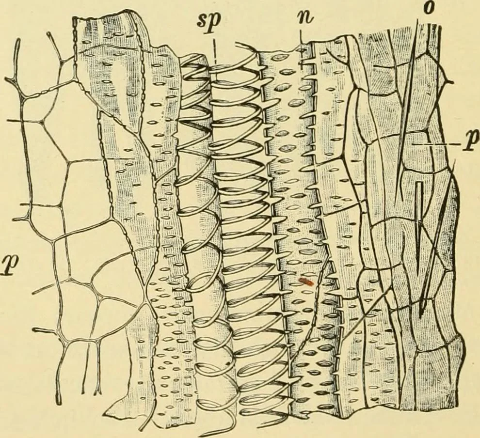
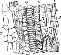
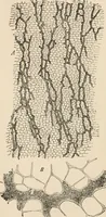
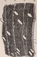
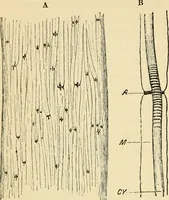
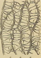
ep Fig. 492. Vanilla. Outer layers of fruit in surface view, ep epicarp with v crystals ofcalcium oxalate; /> parenchyma. X160. (Moeller.) of thin-walled, elongated elements. On the surface between the mem-bers of each pair of placentae, the epidermal and subepidermal layers … Fig. 493. Vanilla. Longitudinal section of fruit flesh, p parenchyma with 0 raphides;5/) spiral vessel; « pitted vessel. X160. (Moeller.) are made up of longitudinal bundles of thread-like mucilaginous cellswhich serve as a conducting tissue for the pollen tubes. They havebeen studied by Busse,…
Plant anatomy · Food adulteration and inspection · Microscopy
Internet Archive · Library of Congress · Public Domain Mark 1.0. Free to use for anything.