Winton, Andrew Lincoln, 1864-1946, Moeller, Josef, 1848-1924 · New York, J. Wiley & sons; [etc., etc.] · leaf 144
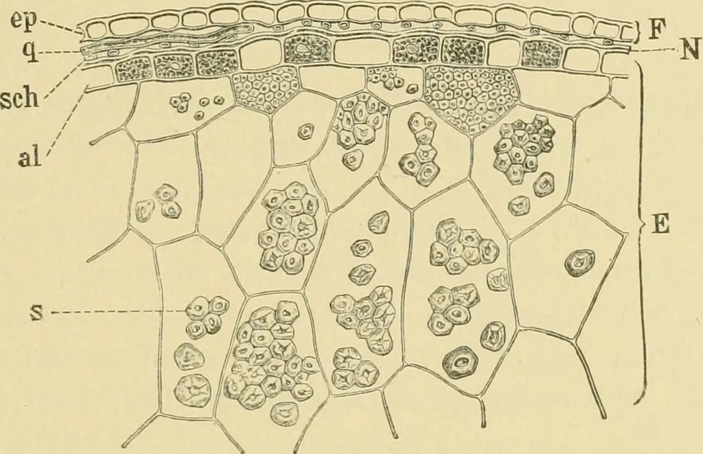
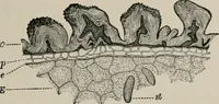
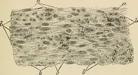
, and more irregular in shape. 3. Tube Cells (sch). These are 2-4 /x wide and often reach the lengthof 300 fl. Perisperm (N). After treatment with alkah, this layer is clearly seenill surface view. The cells arc of large size and have beaded walls. Fig. 89. Green Foxtail. Outer epi-dermis from edge… … FiG. 90. Green Foxtail. Cross section of outer portion of fruit. F pericarp consists ofcp epicarp, q cross cells, and sch tube cells; N perisperm; E endosperm consists ofal aleurone cells and s starch cells. X300. (Winton.) ous hilum fill the parenchyma cells of the endosperm. In the outerlayers…
Food adulteration and inspection · Plant anatomy · Microscopy
Internet Archive · Library of Congress · Public Domain Mark 1.0. Free to use for anything.