Winton, Andrew Lincoln, 1864-1946, Moeller, Josef, 1848-1924 · New York, J. Wiley & sons; [etc., etc.] · leaf 220
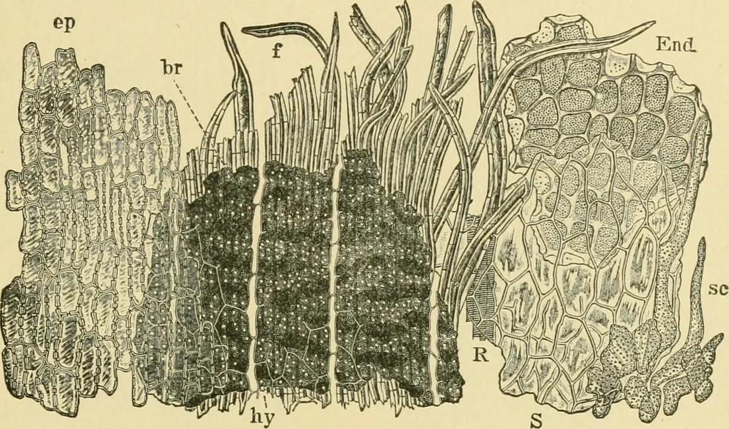
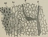
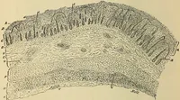
elongated cells,forming wedge-shaped partitions (m). 5. Parenchyma (p). Several rows of partially collapsed parenchymacells form the inner layers of the pericarp. MADIA SEED 199 The Spennoderm (Figs. 158 and 159, S) consists of one distinct layerof parenchyma cells without any striking characters,… … Fig. 159. Madia. Elements of fruit in surface view, ep epicarp; hy hypoderm; brpigment plates; / fiber bundle; S spermoderm with R raphe bundle; sc pitted cellsat base of spermoderm; £H<i endosperm. X160. (Winton.) forms extending in bundles toward the apex. These bundles appearto be distinct…
Food adulteration and inspection · Plant anatomy · Microscopy
Internet Archive · Library of Congress · Public Domain Mark 1.0. Free to use for anything.