Winton, Andrew Lincoln, 1864-1946, Moeller, Josef, 1848-1924 · New York, J. Wiley & sons; [etc., etc.] · leaf 324
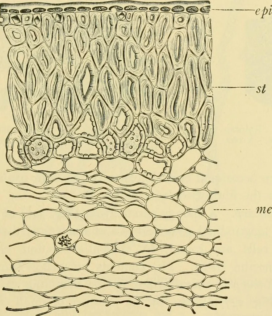
These cells vary in formand are usually between 100 and 200 ix in diameter. The soHtary cellshave thinner walls than those in groups and often contain crystal clustersof calcium oxalate. The bicollateral bundles are accompanied on theouter side by sclerenchyma fibers and rows of crystal chambers.… … mes Fig. 244. Acorn {Quercus sp.). Tissues of shell in cross fection. epi epicarp; st crystalcells and stone ceils; mes mesocarp. (Moellek.) ing a highly characteristic tissue. These cells contain colorless dropsin a brown ground substance. On the upper end in many species arenumerous hairs (Fig.…
Food adulteration and inspection · Plant anatomy · Microscopy
Internet Archive · Library of Congress · Public Domain Mark 1.0. Free to use for anything.
![An illustration from Fundamentum historiae naturalis muscorum frondosorum concernens eorum flores, fructus, seminalem propagationem adjecta generum dispositione methodica. Iconibus illustratis / [Johann Hedwig]. (1782)](https://img.insideoutlibrary.com/img/200/wellcome-wellcome_b28762721-143-1.webp)