Winton, Andrew Lincoln, 1864-1946, Moeller, Josef, 1848-1924 · New York, J. Wiley & sons; [etc., etc.] · leaf 351
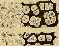
umen. In theinner portion of the cell this mucilaginous wall is thinner, the cavitybeing here bulb-shaped. The thin innermost, or tertiary membrane of 33° FRUIT. the cell-wall greedily takes up safranin, thus bringing out very clearlythe cell cavity, which, taken as a whole, is flask-shaped. 2.… … Fig. 254. Pear. Hairs from suture of endocarp. (Malfatti.) into tube cells such as occur in the apple, is not noticeable, the fiberspassing abruptly into the cross cells of the next layer. 3. Cross Cells, 4. Starch Cells, and 5. Inner Epidermal Cells, alsoPerisperm, Endosperm, Embryo, and Stem are…
Food adulteration and inspection · Plant anatomy · Microscopy
Internet Archive · Library of Congress · Public Domain Mark 1.0. Free to use for anything.