Winton, Andrew Lincoln, 1864-1946, Moeller, Josef, 1848-1924 · New York, J. Wiley & sons; [etc., etc.] · leaf 465
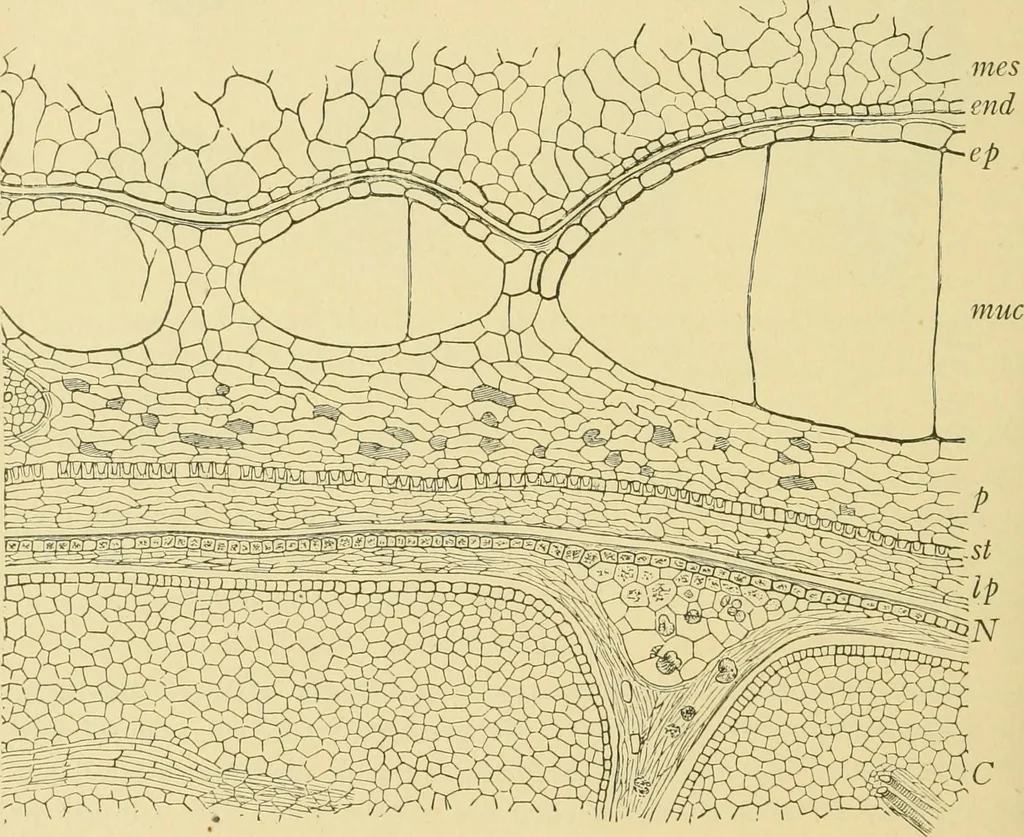
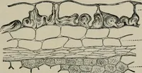
ular spaces. On soaking in water they become slimyand, together with the endocarp, separate easily from the spermoderm. 2. The Endocarp (Figs. 346 and 347, end) is made up of narrow elon-gated cells running transversely or diagonally about the seed and formingthe so-called cross-cell layer. These… … Fig. 346. Cocoa. Cross section of outer portion of bean, mcs inner layers of mesocarp;end endocarp; spermoderm consists of ep outer epidermis, muc mucilage cells, p spongyparenchyma, st stone cells and Ip nutritive layer; A^ perisperm consists of epidermaland obliterated layers; C cotyledon.…
Food adulteration and inspection · Plant anatomy · Microscopy
Internet Archive · Library of Congress · Public Domain Mark 1.0. Free to use for anything.