Winton, Andrew Lincoln, 1864-1946, Moeller, Josef, 1848-1924 · New York, J. Wiley & sons; [etc., etc.] · leaf 609
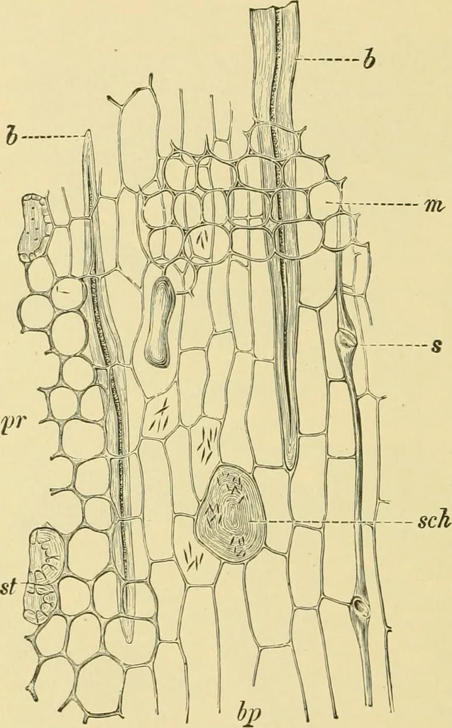
Distributed through this ground tissue are stone cells,many of which are thickened only on the inner side. Both theparenchyma and the stone cells contain rounded starch grains (Fig.505, B) ranging up to 20 n in diameter (mostly over 10 //), with a 588 SPICES AND CONDIMENTS. more or less distinct… … Fig. 504. China Cassia. Radial longitudinal section of bark, pr parench)mia of cortex;bp parenchyma of bast; b bast fibers; st stone cells; sch mucilage cells; 5 sieve tubes;OT medullary rays. X160, (Moeller.) cells are formed, which after a time make up the greater part of the ring.The bast fibers…
Food adulteration and inspection · Plant anatomy · Microscopy
Internet Archive · Library of Congress · Public Domain Mark 1.0. Free to use for anything.