The microscopy of vegetable foods, with special reference to the detection of adulteration and the diagnosis of mixtures (1916)
Winton, Andrew Lincoln, 1864-1946, Moeller, Josef, 1848-1924, Winton, Kate Grace Barber, 1882- · New York, John Wiley & sons, inc.; [etc., etc.] · leaf 614
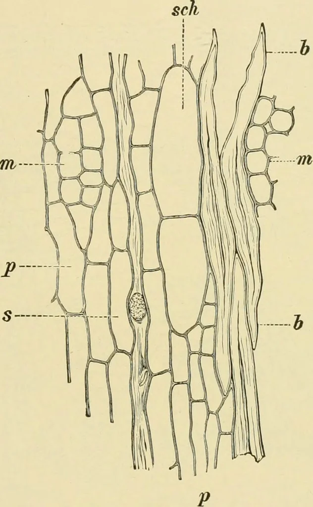
mpound quills*The bark is 1-2 mm. thick, more or less flaky on the outer surface, finelystriate on the inner. It is brittle, and breaks with a smooth fracture.Cross sections examined with the naked eye show a thin yellow outerring and a broad inner zone with yellow dots in a red-brown ground. CLOyE… … Fig. 508. Ceylon Cinnamon. Tangential section of bark, p bast parenchyma; schmucilage cells; b bast fibers; 5 sieve tubes; m medullary rays, X160. (Moeller.) 2. Phelloderm (Fig. 509, P). The inner walls of the cells are stronglythickened and porous. 3. Cortex. The tissue is of parenchyma cells with…
Plant anatomy · Food adulteration and inspection · Microscopy
Internet Archive · Library of Congress · Public Domain Mark 1.0. Free to use for anything.