Sthohr's histology : arranged upon an embroyological basis (1906)
Sthohr, Philipp, 1849-1911, Lewis, Frederic Thomas, 1875 · Philadelphia : P. Blakiston's Son & Co. · leaf 236
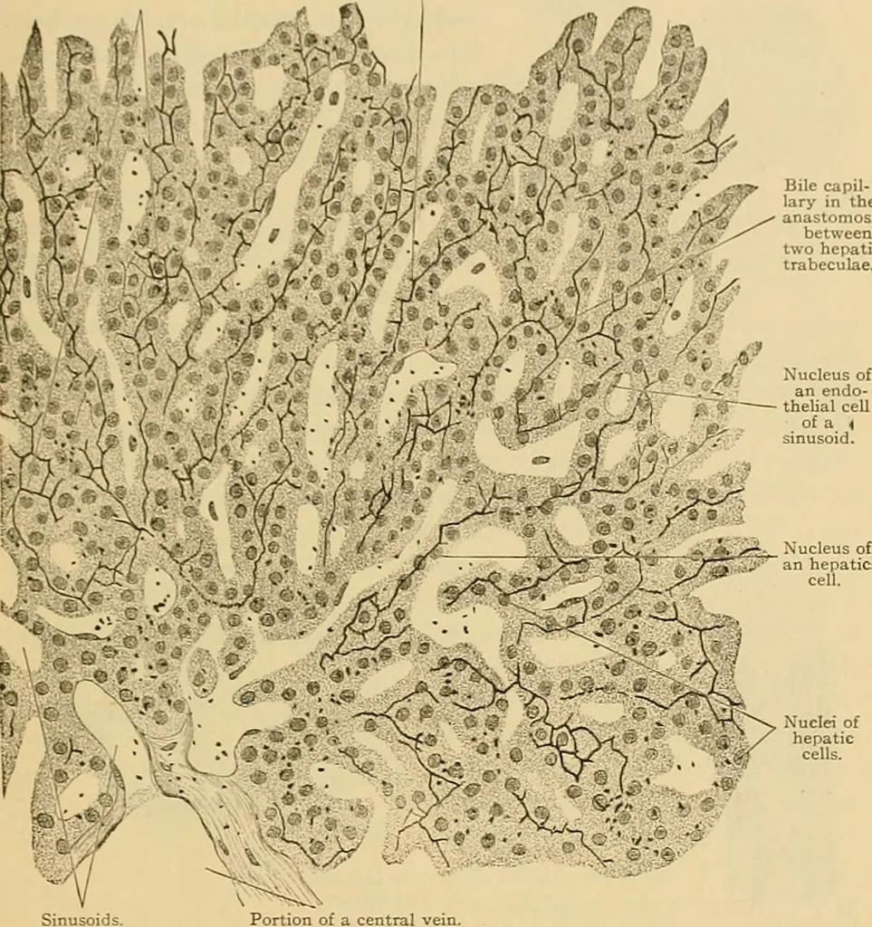
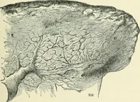
ich they empty are cut longitudinally. X 15. LIVER. beculae as shown in Fig. 254. Near the central veins they form terminalloops. The cells composing the trabeculae are polygonal or cuboidal withan exoplasmic layer which sometimes resembles a cell membrane. Thecells contain round nuclei which are… … Nuclei of hepatic cells. Portion of a central vein Fig. 254.—From a Cross Section of a Human Hepatic Lobule. X 300.Golgi preparation. The boundaries of the hepatic cells could not be seen. The black dots are precipitates of the silver. they may be dark or pale. Often a cell contains two nuclei, and…
Histology · Microscopy
Internet Archive · Public Domain Mark 1.0. Free to use for anything.