Sthohr's histology : arranged upon an embroyological basis (1906)
Sthohr, Philipp, 1849-1911, Lewis, Frederic Thomas, 1875 · Philadelphia : P. Blakiston's Son & Co. · leaf 76
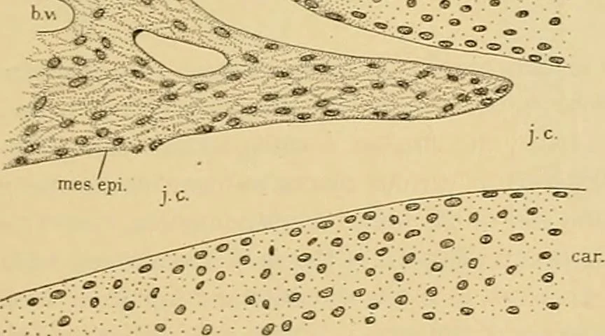
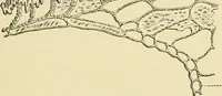
loosein te.xture and a cleft forms in it, containingtissue fluid. This is the joint cavity, Fig. 70.It is bounded by mesenchymal cells whichspread out and form an epithelium, shown inFig. 71. The epithelium may fuse with the articular cartilage so that thelatter, uncovered by perichondrium, forms a… … Fig. 71.—An Enlarged Drawing op the Left Part of the Joint shown in Fig. 70.b. v.. Blood vessel; car., cartilage; j. c, joint ca\-ity; mes. epi., mesenchymal epithelium- ca\it}. Articular cartilages are usually hyaline layers from 0.2 mm. to5 mm. thick, becoming thin at the periphery. The cells…
Histology · Microscopy
Internet Archive · Public Domain Mark 1.0. Free to use for anything.