The microscopy of vegetable foods, with special reference to the detection of adulteration and the diagnosis of mixtures (1916)
Winton, Andrew Lincoln, 1864-1946, Moeller, Josef, 1848-1924, Winton, Kate Grace Barber, Mrs · New York, John Wiley & sons, inc.; [etc., etc.] · leaf 155
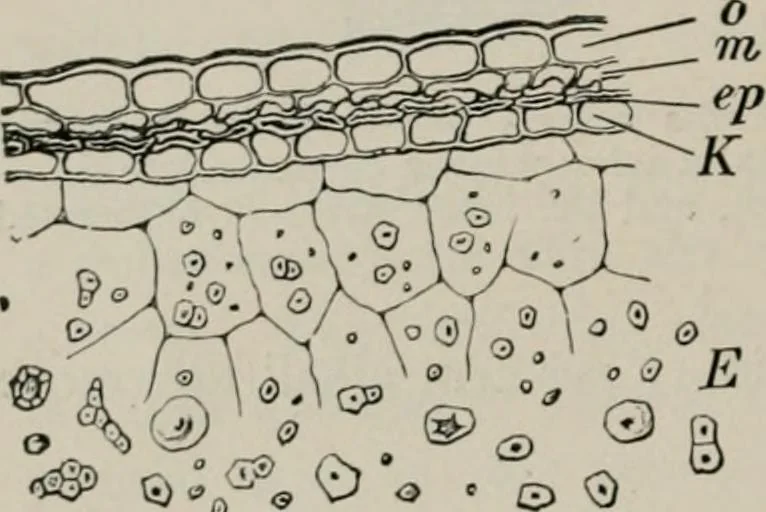
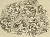
ellular spaces, underlie the ei)i(krmis, and with it form amost valuable means of identification. Greenish or brownish-yellowcell contents render this layer particularly distinct. 3. An Inner Epidermis (ep) of elongated, thin-walled cells, is readilyfound after the addition of cold dilute alkali.… … o ^ (i) _^ ^ . COMMON BUCKIVHEAT. US Surface mounts show that the cells are exceedingly variable both in sizeand wall thickness. 2. Starch Parenchyma (E). Cells of large size with thin walls containthe densely crowded, polygonal starch grains (Fig. io6). Isolated cells