The microscopy of vegetable foods, with special reference to the detection of adulteration and the diagnosis of mixtures (1916)
Winton, Andrew Lincoln, 1864-1946, Moeller, Josef, 1848-1924, Winton, Kate Grace Barber, Mrs · New York, John Wiley & sons, inc.; [etc., etc.] · leaf 250
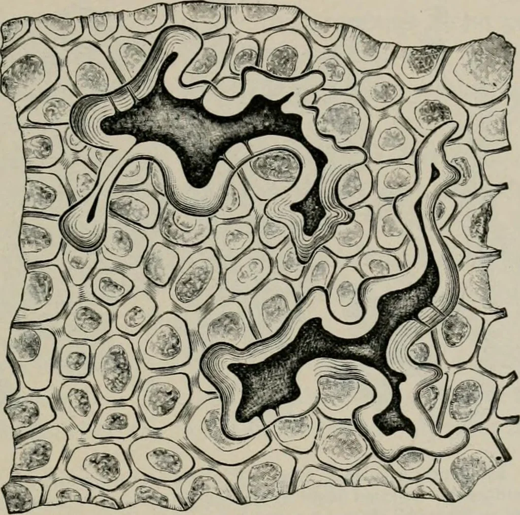
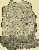
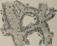
(Fig. 183) consists of thick walled polyg-onal cells about 25 /^ in diameter. This layer, as well as the mesocarp,contains a purple pigment, which, as Hanausek first noted, becomes in-tensely red on addition of concentrated sulphuric acid. 2. The Mesocarp contains so much oil, that a clear idea of… … Fig. 183. Olive {Oka Europea). Epicarp and two stone cells of the mesocarp, seen from beneath. (Moeller.) here and there among this thin-walled tissue are stone cells (Fig. 183)remarkable for their fantastic shapes and especially for the curiousbeaked, T- or Y-shaped excrescences, occurring at the…