The microscopy of vegetable foods, with special reference to the detection of adulteration and the diagnosis of mixtures (1916)
Winton, Andrew Lincoln, 1864-1946, Moeller, Josef, 1848-1924, Winton, Kate Grace Barber, Mrs · New York, John Wiley & sons, inc.; [etc., etc.] · leaf 344
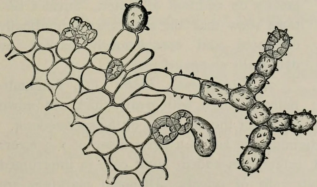
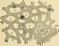
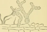
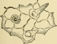
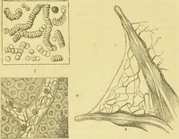
urious, jointed, branching, warty hairs (Fig. 251a)form dead white masses often evident to the naked eye. Some of the Fig. 251. Apple {Pyrus Mains). Isolated elements of thefruit, epi epidermis, hy hypoderm; fruit flesh ele-ments: 5/ stone cells, p^ parenchyma containing amstarch grains, p^ inner… … Fig. 251a. Apple (Pyrus Mains). Hairs from suture ofendocarp. (Malfatti.) 324 FRUIT. hair cells, particularly the terminal ones, are sclerenchymatized, thusfurnishing a distinction from the similar hairs of the pear and quince. Spennoderm. i. The Outer Epidermis (Figs. 2^ib and 25if, ep) isstudied…