Inside Out Library · Explore
Methods in microscopial research; vegetable histology (1905)
Flatters, Abraham · London, Sherratt and Hughes · leaf 134
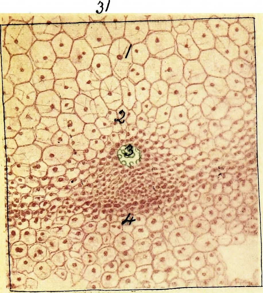 tive budthan above x 30 dia. Fixed as above. Stained with BoraxCarmine and Malachite Green. 1. Central ground tissue. 2. Nodal diaphragm. 3. Secretion cells. 4. Vascularsystem. 5. Lateral bud. 6. Cavity of vascular bundle.7. Growing point of lateral bud. 8. Caniculi in formation. Fig. 34.… … j^
Microscopy · Plant anatomy
Internet Archive · Cornell University Library · Public Domain Mark 1.0. Free to use for anything.
More pictures from Methods in microscopial research; vegetable histology
More like this