Inside Out Library · Explore
Methods in microscopial research; vegetable histology (1905)
Flatters, Abraham · London, Sherratt and Hughes · leaf 152
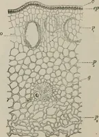
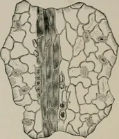
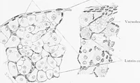
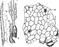
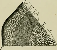
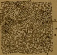
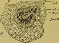
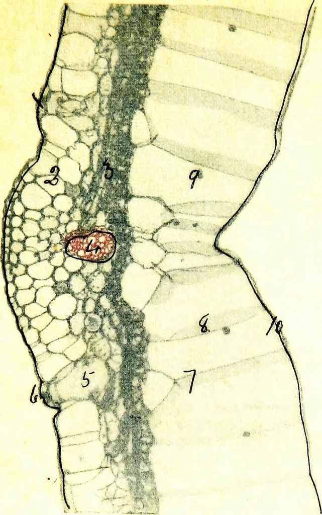 .2. Leaf-trace bundles. 3. Cambium layer. 4. Xylemelements. 5. Primary medullary ray. 6. Central axis ofstem. 7. Stomata bordering the stem cavities. Fig. 44. Epidermis and stinging-hairs from midrib of young Leafof Nettle. Urtica dioicax35 dia. Fixed with Chromo-Acetic solution. Stained with Borax…
Microscopy · Plant anatomy
Internet Archive · Cornell University Library · Public Domain Mark 1.0. Free to use for anything.
More pictures from Methods in microscopial research; vegetable histology
More like this