The microscopy of vegetable foods : with special reference to the detection of adulteration and the diagnosis of mixtures (1906)
Winton, Andrew Lincoln, 1864-1946, Moeller, Josef, 1848-1924 · New York : John Wiley & Sons · leaf 559
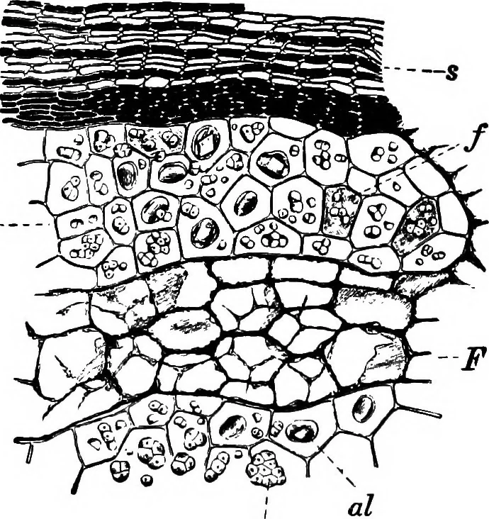
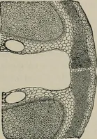
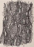
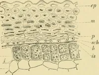
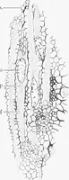
l portion. As seen in cross-section, the cells of both palisadelayers are wavy in outline, owing to the irregularities of the surface ofthe seed, formed by the pressure of the mace during growth. Primary Perisperm (Fig. 453, qjs). 1. Fiber Layer. Tschirch andOesterle have shown that the fibers of… … am Fig. 4S4. Nutmeg. Cross section of kernel, sprimary perisperm; F secondary perisperm ofveins; B endosperm with am starch grains, alaleurone grains and / pigment cells. X160. (MOELLER.)
Plant anatomy · Food adulteration and inspection · Microscopy
Internet Archive · Cornell University Library · Public Domain Mark 1.0. Free to use for anything.