The microscopy of vegetable foods : with special reference to the detection of adulteration and the diagnosis of mixtures (1906)
Winton, Andrew Lincoln, 1864-1946, Moeller, Josef, 1848-1924 · New York : John Wiley & Sons · leaf 91
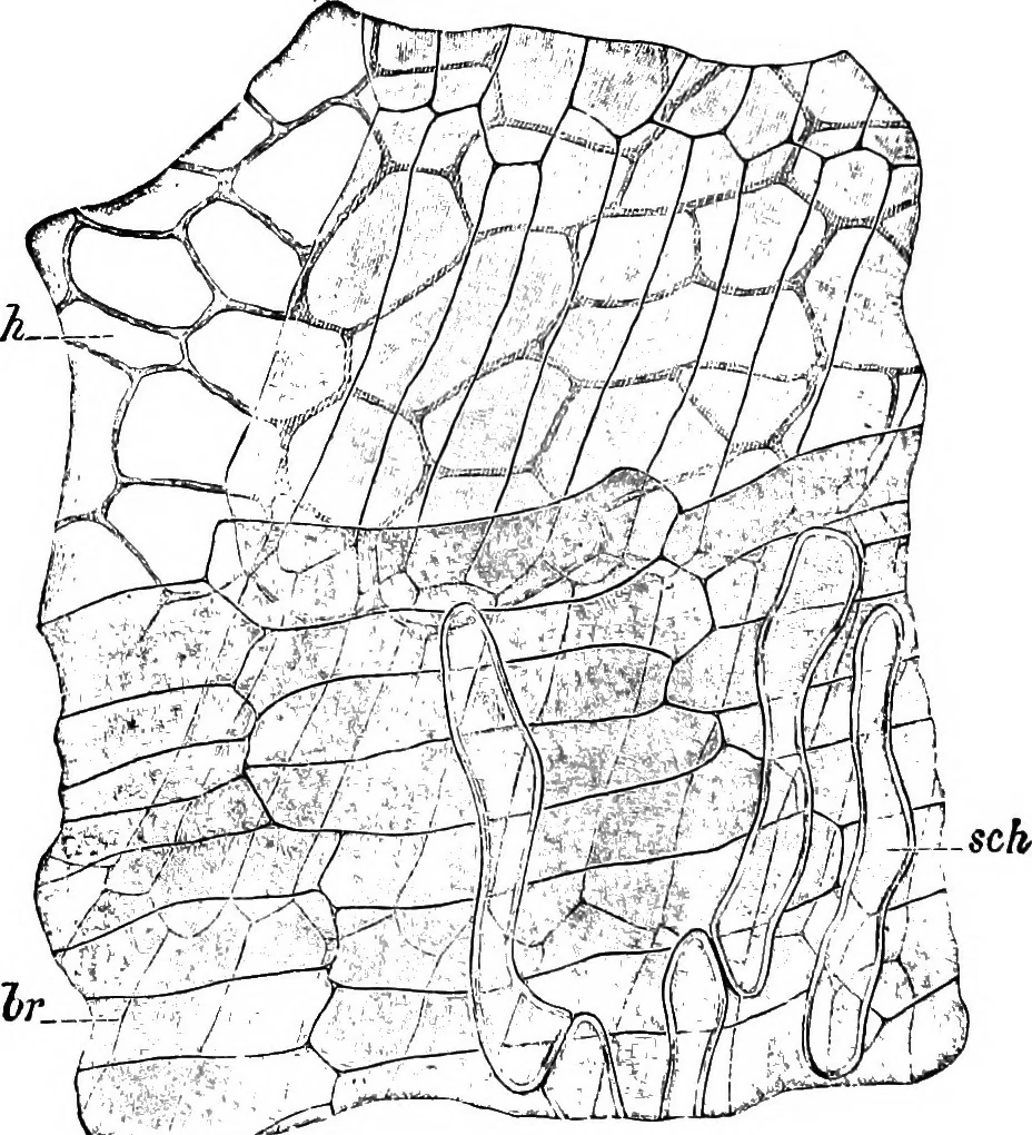
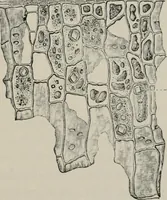
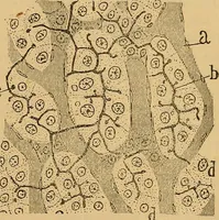
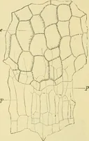
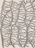
sections of these cells are circular or elliptical. Spermoderm (Figs. 33 and 37, br). In cross-section, before treat-ment with reagents, the two layers of the spermoderm appear like yellow-brown, structureless membranes, the inner somewhat darker than theouter; but on treatment with Javelle water,… … Fig. 37. Wheat. Surface view of sch tube cells, br two crossing layers of the spermodennand/i perisperm. X300. (Moellee.) Perisperm (Figs. 3;^, 37 and 38, h). The remains Q-f the nucellus orbody of the ovule, known as the nucellar layer, and by some authorsbecause of its colorless, almost…
Plant anatomy · Food adulteration and inspection · Microscopy
Internet Archive · Cornell University Library · Public Domain Mark 1.0. Free to use for anything.