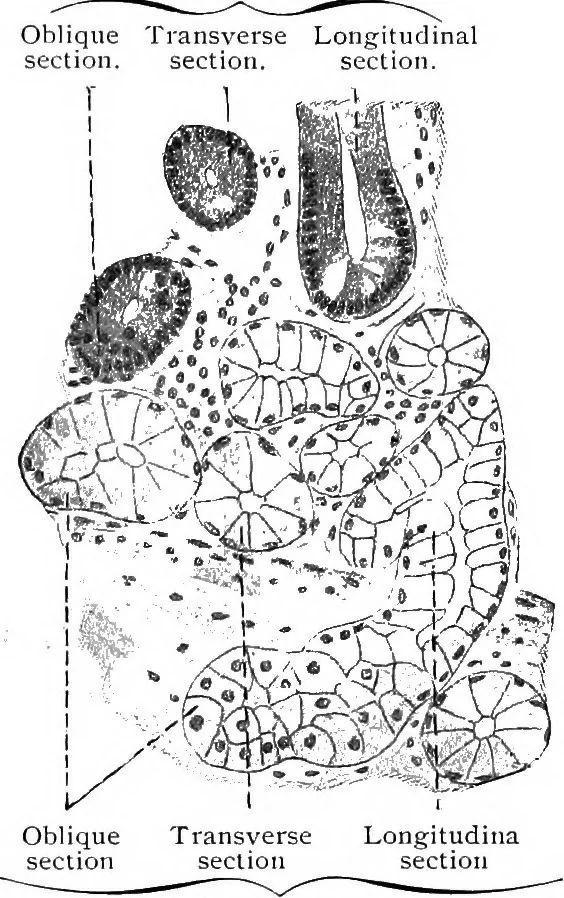
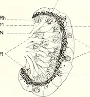
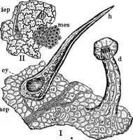
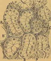
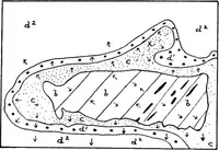
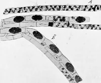
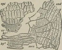
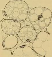
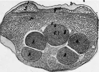
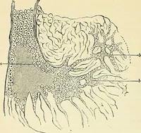
connective tissue, the tunica propria (Fig. 231). There are other andlarger elevations in the lining of the small intestine, known as circular folds(plicae circulares) [valvulae conniventes]. As shown in Fig. 232, theirinterior is formed by the submucosa and their surface is covered by theentire… … of the tubules of a duodenal gland. Fig. 230.—From a Section of a HumanDuodenum. X 240. Only the lower half of the mucosa and upperhalf of the submucosa are sketched. Alarge portion of the duodenal gland liesabove the muscularis mucosae. Digitized by Microsoft® 2o6 HISTOLOGY. (Fig. 229), and beyond…
Histology · Microscopy
Internet Archive · Cornell University Library · Public Domain Mark 1.0. Free to use for anything.