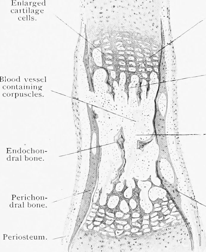
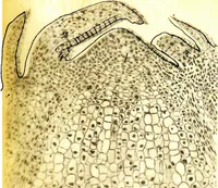
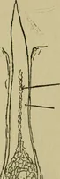
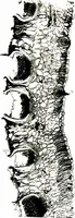
is in part deposited in contact with cartilagein the following manner. Fig. 65 shows a longitudinal section of a developing phalanx. Oneither side of the shaft a strip of bone is seen, formed from undifi:erentiatedcells of mesenchymal origin, situated in the perichondrium. It is calledperichondral… … Calcifiednialrix ofcartilage. Spicule ofcalcifiedmatrix. Marrowspace. Pcrichon-■ dral bone. Fic. 66—FrO-M a DoRso-r.ALMAR Longitudinal Section ok aMiddle-finger Phalan.\ of a Hitman Ff.tus Four MonthsOld. X 60. Digitized by Microsoft® 62 HISTOLOGY. one lacuna. The lacunae also have increased in…
Histology · Microscopy
Internet Archive · Cornell University Library · Public Domain Mark 1.0. Free to use for anything.