The microscope; an introduction to microscopic methods and to histology (1899)
Gage, Simon Henry, 1851-1944 · Ithaca, N. Y., Comstock publishing company · leaf 47
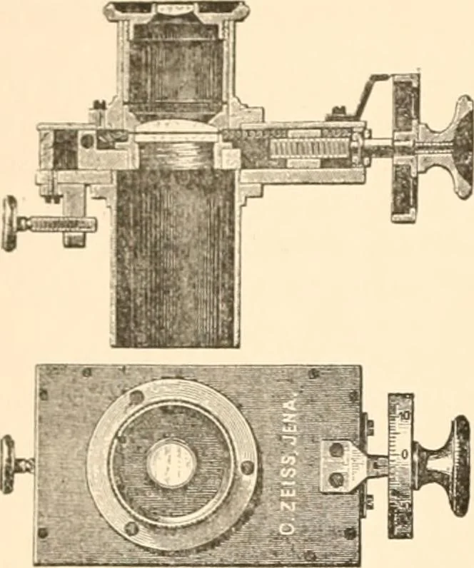
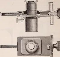
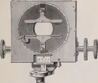
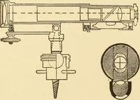
Fig. 33. fig. 34. Figs. 33-34 Ocular Micrometer with movable scale. Fig. 33 is a tide \the ocular while Fig. 34. gives a sectional end view, and shows the ocular mitei in position. In both the screw which moves the micrometer i tat the left. {From Bausch & Lomb Opt. Co.) 26 MICROSCOPE AND… … Fig. 35. Ocular Screw-Micrometer 7vithcompensation ocular 6. The tipper figureshows a sectional view of the ocular and thescrew for moving the micrometer at the right.At the left is shown a clamping screzv tofasten the ocular to the upper part of the mi-croscope tube. Below is a face vieza,…
Microscopy -- Technique
Internet Archive · Public Domain Mark 1.0. Free to use for anything.