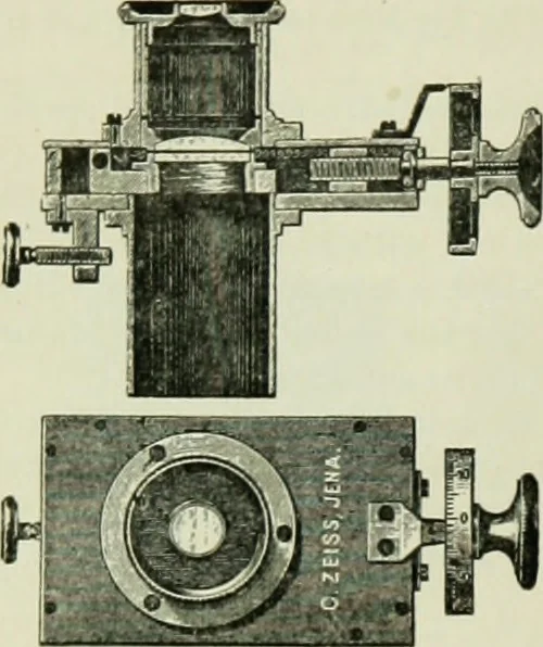
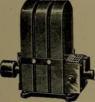
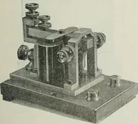
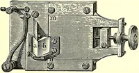
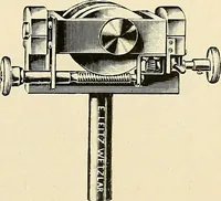
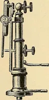
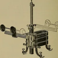
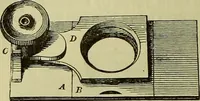
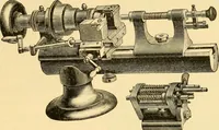
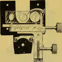
Fig. 33Figs. 33-34 Ocular Micrometer with movable scale. Fig. 34. Fig. 33 w a side view of the ocular while Fig. 34 %iv?s a sectional end view, and shows the ocular microme-ter in position. In both the screw which moves the micrometer is shown at theleft. (From Bausch & Lomb Opt. Co.) 26… … Fig. 35- Ocular Scrcu-Micrometer withcompensation ocular 6. The upper figureshows a sectional view of the ocular and thescrew for moving the micrometer at the right.At the left is shown a damping screw tofasten the ocular to the upper part of the mi-croscope tube. Below is a face view, showingthe…
Microscopy
Internet Archive · Medical Heritage Library · Public Domain Mark 1.0. Free to use for anything.