The microscopy of vegetable foods, with special reference to the detection of adulteration and the diagnosis of mixtures (1916)
Winton, Andrew Lincoln, 1864-1946, Moeller, Josef, 1848-1924, Winton, Kate Grace Barber, 1882- · New York, John Wiley & sons, inc.; [etc., etc.] · leaf 388
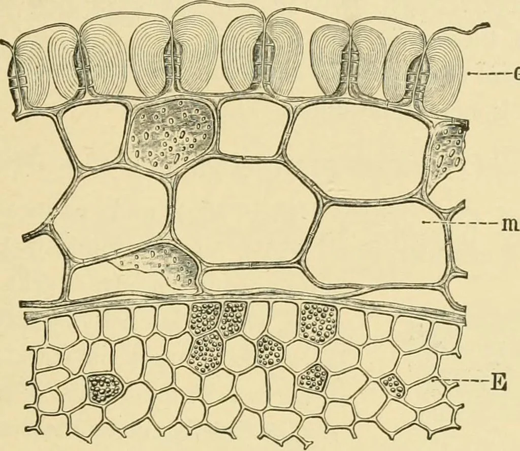
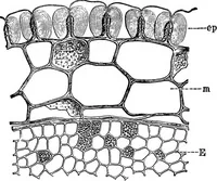
and radial walls are sclerenchymatously thickened (doublewalls often 20 /t), and in addition the radial walls and sometimes theouter and inner walls have a transparent mucilaginous layer of distinctlystratified structure which nearly fills the cell cavity. Treated with chlor-zinc iodine the… … Fig. 285. Cultivated Cranberry. Seed in cross section, ep epidermis of spermodermwith sclerenchymatized and mucilaginous layers; m inner spermoderm; E endospermX160. (WiNTON.)
Plant anatomy · Food adulteration and inspection · Microscopy
Internet Archive · Library of Congress · Public Domain Mark 1.0. Free to use for anything.
![An illustration from Fundamentum historiae naturalis muscorum frondosorum concernens eorum flores, fructus, seminalem propagationem adjecta generum dispositione methodica. Iconibus illustratis / [Johann Hedwig]. (1782)](https://img.insideoutlibrary.com/img/200/wellcome-wellcome_b28762721-143-1.webp)