Winton, Andrew Lincoln, 1864-1946, Moeller, Josef, 1848-1924 · New York, J. Wiley & sons; [etc., etc.] · leaf 275
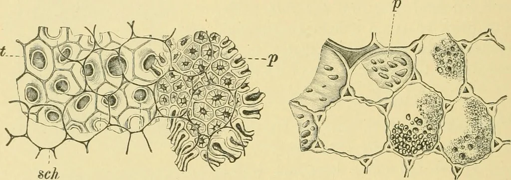
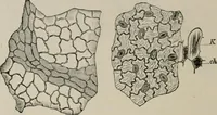
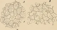
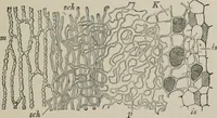
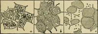
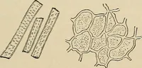
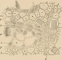
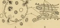
radial walls and the edges of the tangential walls. The remainder of the cotyledons consists of isodiametric cells withmuch swollen, porous walls, often 15-25 n thick. This thickening isespecially marked at the angles. Ovoid aleurone grains up to 20 /i, oftencontaining large crystalloids, are the… … Fig. 203. Yellow Lupine. Elements of spermo- Fig. 204. Yellow Lupine. Collen- derm in surface view, p palisade cells; / hour- chyma cells of cotyledon with aleu- glass cells ^subepidermal layer) showing contour rone grains. /> porous wall. X300. of base and constriction; sch spongy paren-…
Food adulteration and inspection · Plant anatomy · Microscopy
Internet Archive · Library of Congress · Public Domain Mark 1.0. Free to use for anything.