Winton, Andrew Lincoln, 1864-1946, Moeller, Josef, 1848-1924 · New York, J. Wiley & sons; [etc., etc.] · leaf 628
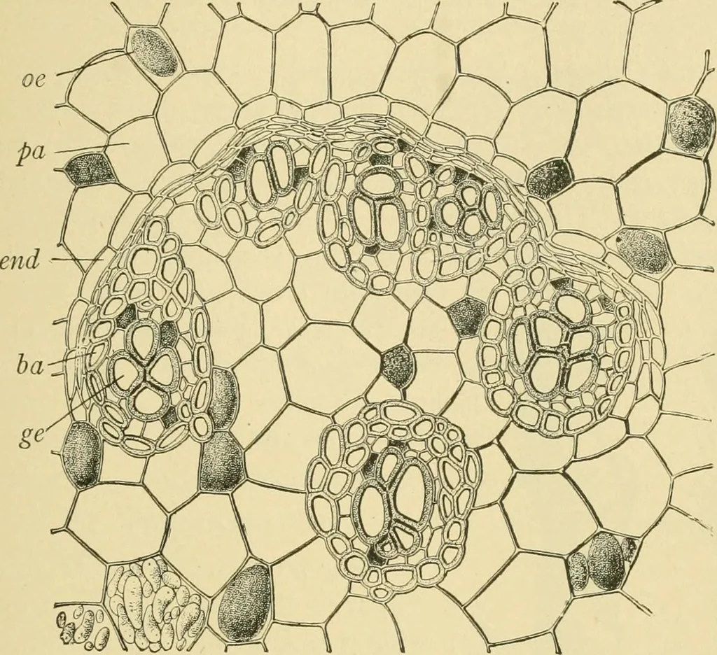
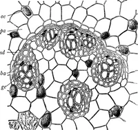
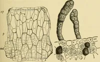
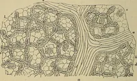
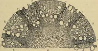
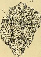
Fig. 520. Galangal {Alpinia officinarum). Outer layers of rhizome in surface view, epepidermis; rp brown parenchyma; g tannin grains; h oil cell. Xi6o. (Moeller.) lumps, but no starch. Further inward the cells have thick porous walls,and contain starch grains. 3. Endodermis (Fig. 521, end). The… … Fig. 521. Galangal. Cross section of rhizome, pa parenchyma, oe oil cells, and end endo-dermis of the corte.x; ha bast fibers and ge vessels of a bundle. (GiLG.)
Food adulteration and inspection · Plant anatomy · Microscopy
Internet Archive · Library of Congress · Public Domain Mark 1.0. Free to use for anything.