Winton, Andrew Lincoln, 1864-1946, Moeller, Josef, 1848-1924 · New York, J. Wiley & sons; [etc., etc.] · leaf 90
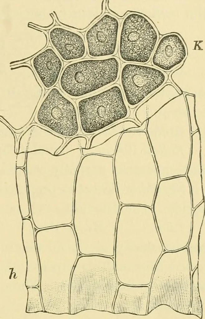
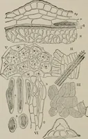
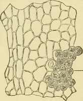
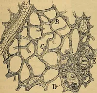
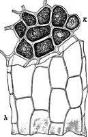
er.) Perisperm (Figs. 33, 37 and 38, h). The remains of the nucellus orbody of the ovule, known as the nucellar layer, and by some authorsbecause of its colorless, almost structureless appearance, as the hya-line layer, can be seen in surface preparations only under the mostfavorable conditions. To… … tion of the inner skin, which may be readily separated after this treat-ment, and gently press sidewise with acover-glass. If zinc chloride iodine isnow added, both cell layers of the spermo-derm are colored brown, the perispermand the remaining coats blue. Endosperm, i. The Aleurone Layer(Figs.…
Food adulteration and inspection · Plant anatomy · Microscopy
Internet Archive · Library of Congress · Public Domain Mark 1.0. Free to use for anything.