The microscopy of vegetable foods : with special reference to the detection of adulteration and the diagnosis of mixtures (1906)
Winton, Andrew Lincoln, 1864-1946, Moeller, Josef, 1848-1924 · New York : John Wiley & Sons · leaf 454
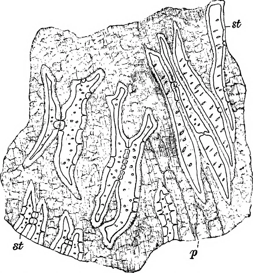
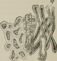
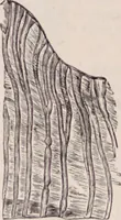
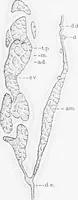
Fig. 336. Coffee. Sclerenchyma fibers of endocarp. X160. (Moeiler.) and may be readily removed in one piece after soaking the seed for somehours in water. I. Sclerenchyma Cells (Fig. 333, 8; Fig. 337, st) form the character-istic outer layer. In the early stages of development the coat is uninter- … Fig. 337. CoSee. Spermoderm in surface view, st sclerenchyma; p compressed paren-chyma. X 160. (MOELLEE.) rupted, but in the mature seed, as a result of more rapid growth of adjoin-ing tissues, they arc more or less detached, occurring singly, in pairs or 432 ALK/tLOID/IL PRODUCTS. in groups,…
Plant anatomy · Food adulteration and inspection · Microscopy
Internet Archive · Cornell University Library · Public Domain Mark 1.0. Free to use for anything.