The microscopy of vegetable foods : with special reference to the detection of adulteration and the diagnosis of mixtures (1906)
Winton, Andrew Lincoln, 1864-1946, Moeller, Josef, 1848-1924 · New York : John Wiley & Sons · leaf 307
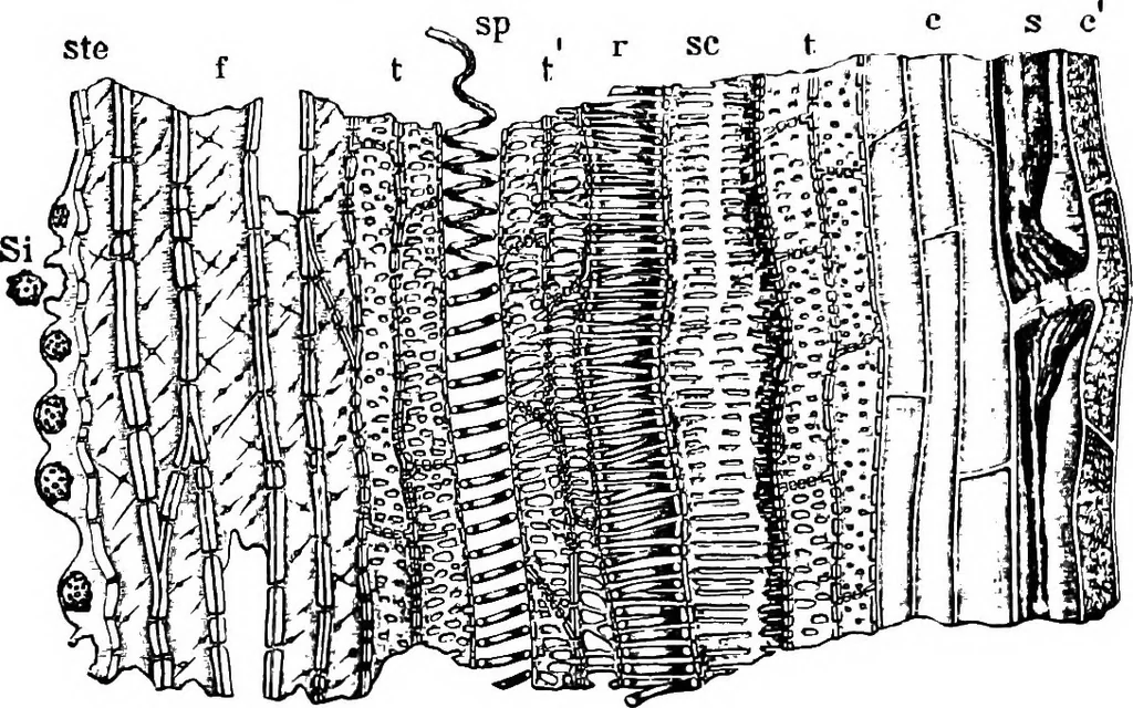
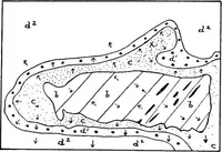
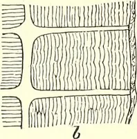
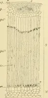
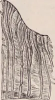
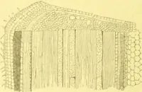
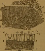
heath of bast fibers; ph two phloem groups; x xylem; p parenchyma of groundtissue; a rudimentary bundle belonging to sriiall branch. X90. (WiNTON.) 2. Mesocarp. The outer portion consists of a ground tissue of thick-walled, porous cells, through which pass longitudinally arranged strands ?84 NUTS.… … Fig. 232. Cocoanut. Longitudinal section of a large (mesocarp) fiber, ste stegmata;Si silicious body; / bast fibers; t tracheids with small pits; t tracheids with large pits;i^ spiral vessel; r reticulated vessel; jc scalariform vessel; j sieve tube; c and ccambi-form cells. X300. (Winton.) inner…
Plant anatomy · Food adulteration and inspection · Microscopy
Internet Archive · Cornell University Library · Public Domain Mark 1.0. Free to use for anything.